Print This Page

Proximal Interphalangeal Joint (PIP Joint)
- The phalangeal head has a groove and the phalangeal base has a ridge and they contribute to joint stability and limit motion to flexion/extension
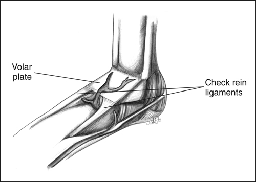
- Check-rein ligaments are the longer proximal attachment of the volar plate and tighten as the middle phalanx extends and limits hyperextension
- The volar plate is the attachment for the A3 pulley which stabilizes the flexor tendons and is essential for digit function
- The PIP joint is stabilized dorsally by the central band and triangular membrane and laterally by the lateral bands and retinacular ligaments
Transverse Retinacular Ligaments
- Anatomy: originates on the flexor tendon sheath at the PIP and inserts on the lateral edges of the conjoined bands
- Function: prevents excessive dorsal shift of the lateral bands when the PIPJ extends and when in flexion they assist with pulling the lateral bands volarly over the PIPJ
- Pathology: attenuation of these ligaments leads to excessive dorsal position of the lateral bands and ultimately a swan neck or with a contracture of the transverse retinacular ligament and attenuation of the triangular ligament will lead to translation of the lateral bands and a boutonniere deformity.
Oblique Retinacular Ligament: (might also see it termed oblique retinacular ligament of Landsmeer)
- Anatomy: the ORL originates from the proximal phalanx and then inserts into the terminal extensor tendon crossing the collateral ligaments
- Function: It links the motion of the DIP and PIP
- Pathophysiology: May be involved with Dupuytren's and contracture of it may contribute to displacement of the lateral bands and boutonniere injury
- Test for ORL tightness: When the ORL is tight it does not allow for passive DIPJ flexion with the PIPJ passively extended; conversely if the PIPJ is in flexion the DIP can flex
Volar Plate:
- Function: prevent hyperextension
- Anatomy: thickening of joint capusule volar to the MP joint; origin is the head of the metacarpal and insertion is via the checkrein ligaments
- Biomechanics: loose in flexion and tight in extension
True or False: The critical corner of the PIPJ is where the accessory and proper collateral ligaments, and volar plate all converge at the middle phalanx.
